Events / Education
International Cancer Imaging Society Meeting and 18th Annual Teaching Course
Sun 07 Oct 2018 Tue 09 Oct 2018 Society
On the French Riviera between Monte Carlo and San Remo.
Advancing Cancer Imaging: Improving Patient Outcomes
High quality educational lectures: refresh your knowledge of cancer imaging and learn about cutting-edge developments
• Listen to state of the art multimodality cancer imaging (MRI, CT and PET)
• Bring your knowledge into practice in workstation based teaching in our hands-on workshops
• Focus on interventional radiology in oncology: from diagnosis to treatment
• Experience the role of cancer imaging in multidisciplinary interaction: treatment selection, early response prediction and treatment adaptation
• Learn how big data initiatives are changing the future of cancer imaging: radiogenomics, machine learning, AI
• Interact with hematologists in a special session on bone marrow imaging (in conjuction with the International Workshop on PET in Lymphoma and Myeloma)
In addition:
• Presentations of cases you would like to read again
• Proffered papers (oral, poster and case presentations)
In Collaboration with the International Workshop in Lymphoma and Myeloma
![]()
Supported by: The French Society of Radiology
President: Prof. Wim Oyen, London, UK
Programme Committee: Prof. Wim Oyen (UK), Stefan Diederich (DE), Aslam Sohaib (UK), Prof Harriet Thoeny (CH), Olivier Lucidarme (FR), Giovanni Morana (IT)

Venue: Palais de L’Europe, 8 Avenue Boyer, 06506 Menton, France
Date: Sunday 7th to Tuesday 9th October 2018
Keynote lectures:
Big data initiatives changing the future of cancer imaging and therapy
PSMA molecular imaging and theranostics
Cancer imaging: through a looking glass
Main sessions:
Myeloma
Lymphoma and myelofibrosis
Imaging musculoskeletal cancer
Imaging immunotherapy
Clinical needs in pancreatic tumours
Liver tumours I
Lung cancer
Federation du cancer from the French Society of Radiology
Multiparametric imaging
Liver tumours II
Break-out sessions:
Abdominal oncology
Interventional radiology
Neurooncology
Imaging complications of cancer therapy
Haematologic cancer
Cases I would like to read again
Computer hands-on workshops:
Lung cancer staging
Pancreatic lesions
Whole body MRI
Prostate cancer
Gynaecological cancer
HCC diagnostic using LIRADS with CT, MRI or CEUS?
Scientific paper sessions and poster exhibition
Prizes for the best oral presentations and best posters will be awarded at the course party on Monday evening.

Other info:
Course party at the Casino, Menton. Monday 8th October 19.30 €50 for delegates and Fellows' partners & guests (unlimited number of tickets can be purchased). 1 Complimentary ticket for Fellows.
Fellows' AGM on Saturday 6th October 16.30 to 17.30
Fellows' dinner at Maison Martin et Fils, 7 Rue de Marins, Menton. Saturday 6th October 19.00. Complimentary limited to two tickets per fellow.
Travel Documents
Things to Do in MentonAccommodation
Delegates are responsible for making their own arrangements for accommodation and should book via the given web link. We strongly recommend you book early to secure the best rates.
The top 10 rated hotels in Menton (starting from £42 per night at time of writing) - https://www.tripadvisor.co.uk/Hotels-g187231-Menton_French_Riviera_Cote_d_Azur_Provence_Alpes_Cote_d_Azur-Hotels.html
Getting to Menton
By Air
Nice Côte d'Azur is the closest international airport, 35km away or a 45 minute drive.
By Taxi
You can prebook a taxi (from €70 per car), or share the journey and get a larger 8 seater minibus (from as little as €90 per van) by clicking here.
By Train
To travel to Menton Station from Nice Airport please take a taxi to Nice Rail Station, and then a direct train from €5.50 one way. You may book these tickets here 12 weeks before the date of travel.
If you are travelling by train from a different part of France, click here to see an interactive map of the French Railway network.
By Bus
It is possible to travel to Menton by bus, however we do not recommend this as the train is excellent value and much quicker. If you would prefer to take the public bus, please board bus number 110 from Nice Airport. Not all of these are direct and so you may have to change at Monaco.

You will be able to generate your own certificate of attendance once you have completed our daily online feedback form.
The Royal College of Radiologists (Category 1 (external) CPD credits)
Sunday 7 October - 6.5 credits
Monday 8 October - 6.5 credits
Tuesday 9 October - 4 credits
European Accreditation Council for CME (EACCME)
(European CME credits (ECMEC)) have been applied for
American delegates can have their EACCME European CME Credits converted to AMA PRA Category 1 Credit™ by applying to the AMA.
Download the 1 page from here:- https://www.ama-assn.org/sites/default/files/media-browser/public/cme/eaccme-application.pdf
Send the completed form to the AMA together with the EACCME certificate* and the non-refundable processing fee (AMA members $30 and non AMA members $75). AMA certificates will be emailed within 3 business weeks.
* The EACCME certificate will be automatically emailed to you once you have completed the ICIS course daily electronic surveys.
Please visit this link for further information https://www.ama-assn.org/education/uemseaccme-cme-credit-recognition
» Guidelines » Examples » Download Abstract Guidelines
General Rules
Abstracts that do not adhere to the following important points will be rejected:
> The title should not exceed 15 words. The title should be in bold, sentence case with no full stop at the end and no underlining.
> The abstract should not exceed 250 words.
> Please use authors’ initials and surnames only. No full stop at the end. Underline the name of the corresponding author. A comma should separate author names. Where authors are from a number of different institutions, the appropriate institution number from the affiliation list should be given as a superscript number immediately after each author's name, e.g.: John Smith1, Susan Jones1, Bill Fisher2. An asterisk * should be used to link the corresponding author with their email address
> Affiliations should include institute, town and country. Where there are multiple affiliations, each should be listed as a separate paragraph. Each institute should appear in the order used against the author names (see above paragraph) and show the appropriate superscript number, e.g.:
1 University, Town, State, USA
2 University, Town, UK
2 Company, Town, Country
> Qualifications should be omitted.
> Do not include references, tables or figures.
> Please do not use block capitals.
Main text
> In structured abstracts, paragraph headings should be typed in bold with no colon at the end. Do not use the heading ‘Abstract’. Each heading should be in a separate paragraph.
Aim
Followed by regular text, on a new line and in the same format as shown above for main text.
Materials & Methods
Results
Conclusions
Consent to publish If the abstract contains details relating to individual participants (for example a case report), written informed consent for the publication of these details must be obtained from the participants and a statement to this effect should appear at the end of the abstract. Our guidelines for consent statements can be found here: http://www.biomedcentral.com/about/editorialpolicies#Ethics. If the patient is deceased consent for publication should be obtained from the next of kin and if the patient is under 16 consent should be obtained from the parent or guardian.
Please find examples at the bottom of the page.
You may choose among four different abstract types:
> Oral Scientific Presentation
> Oral Case Based Presentation
> Poster Scientific Presentation
> Poster Educational Presentation
Oral Scientific Presentation
The abstract should be separated into “Aim”, “Methods”, “Results” and “Conclusion”. The abstract limit is 250 words. Abstracts should not include promissory notes such as “We will provide additional data during our presentation.” Authors of accepted oral presentations will be invited for a presentation within the Scientific Paper Sessions. Presentation time will be 8 minutes with 2 minutes for Q&A (depending on the final program).
Oral Case Based Presentation
The abstract should have an image and three teaching points. The abstract limit is 250 words. Abstracts should not include promissory notes such as “We will provide additional data during our presentation.” Authors of accepted oral case based presentations will be invited for a presentation within the Scientific Paper Sessions. Presentation time will be 8 minutes with 2 minutes for Q&A (depending on the final program).
Poster Scientific Presentation
The abstract should be separated into “Aim”, “Methods”, “Results” and “Conclusion”. The abstract limit is 250 words.
Poster Educational Presentation
The abstract should be separated into “Learning Objectives”, “Content Organisation”, and “Conclusion”. The abstract limit is 250 words.
Proceedings of the 18th International Cancer Imaging
Abstracts selected for presentation will be published in the Proceedings of the 18th International Cancer Imaging, given to all delegates and faculty. A downloadable version of your submission will be made available to our membership on the members only area of our website, and be available on the Cancer Imaging open access website. Authors of selected abstracts will be notified after Friday 6th July 2018.
It will be obligatory for all scientific presenters to be members of ICIS at the time of presentation in Menton. The annual membership fee of €95 will be added to the scientific presenters’ fee at registration if current membership is not in place. Please note that we have a discounted trainee membership at €39 and a much reduced trainee registration fee for the course. Membership will run for one year from date of registration; all standard member benefits will apply.
Financial help will be available for junior proffered presenters. This will be decided on application and on a case-by-case basis. Kindly supported by the French Society of Radiology.
Applicants for financial aid please email [email protected] for further information.
Queries may be addressed to the ICIS Secretariat
Tel: +44 (0) 7956 814964 or Email: [email protected]
Submission deadline: Monday 4th June 2018
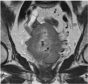
EXAMPLE – ORAL CASE BASED PRESENTATION
Diagnosis: Advanced Cervix Cancer
Sala E.
Memorial Sloan Kettering Cancer Center, New York, USA
Images:
Teaching/Discussion points:
> Discuss the role of MRI in treatment slection and planning of patients with cervical cancer
> Describe MRI Features of parametrial and pelvic sidewall invasion
> Highlight potential pitfalls
EXAMPLE - POSTER EDUCATIONAL
Chemotherapy induced cardiomyopathy: an overview, imaging features, and future prospective
Firstname A Lastname1*, Firstname B Lastname2, Firstname C Lastname3
1University, Town, State, USA
2University, Town, UK
3Company, Town, Canada
*Email address of corresponding author if being included
Learning objectives:
To review the spectrum of imaging findings of chemotherapy- induced cardiomyopathy in correlation with most common cytotoxic drugs and regimens.
Content organisation:
Cardio toxic effect of chemotherapy is a well-recognized problem in cancer patients. Cardio toxicity depends on multiple predisposing factors, specific components of the chemotherapy regimen, length of treatment, and dosage.
We will present the spectrum of most common cardiotoxic chemotherapy agents and their combinations, specific effects on the myocardium, and imaging features of cardiomyopathies induced by chemotherapy.
We will review pathophysiology of chemotherapy induced cardiomyopathy including:
- Dose dependent cardiomyopathy
- Predisposing conditions –diabetes, presence of coronary artery disease, age.
- Potential reversibility
We will discuss imaging characteristics of chemotherapy induced cardiomyopathy
- Imaging modalities ( Echocardiography, Cardiac MR, and MUGA)
- Importance of monitoring cardiac function during and after treatment
- Distribution of late Gadolinium enhancement (LGE)
- Emerging technologies for early diagnosis of cardiomyopathy in cancer patients
Conclusions:
Chemotherapy induced cardiomyopathy is a common problem among cancer patients, increasing long term morbidity and mortality and often leading to disability. Patients receiving chemotherapy treatment, particularly cardio toxic agents, should be routinely assessed for cardiac function to diagnose cardiomyopathy during the early phase of treatment and to prevent development of irreversible heart failure.
EXAMPLE POSTER SCIENTIFIC
The value of 68Ga-PSMA enhanced MR-PET in patients with biochemical recurrent prostate cancer
Firstname A Lastname1*, Firstname B Lastname2, Firstname C Lastname3
1University, Town, State, USA
2University, Town, UK
3Company, Town, Canada
*Email address of corresponding author if being included
Aim: In patients with prostate Cancer increased levels of PSMA can be measured. Recently a new tracer, 68Ga-PSMA, was developed as a specific marker for hybrid imaging (PET/CT, MR-PET). In this study we evaluated the accuracy of 68Ga-PSMA in patients with rising PSA after radical prosatectomy, so called „biochemical recurrent prostate cancer“ (BRPC).
Materials and Methods: A total of 322 patients with BRPC underwent a MR-PET examination (Siemens Biograph mMR) after injection of about 150 mBq 68Ga-PSMA. Images were evaluated in cosensus by one experienced nuclear medicine physician and one radiologist. Pelvine lymphnode dissection was performed in most of the patients according to a predefined template with 8 fields. Lymphnode involvement was evaluated according to a 5 point scale with a patient- and a field-based analysis. These findings were startified according to PSA-values.
Results: Four patients were excluded from the study for different reasons. Sensitivity for detction of recurrence was 95.7 % for PSA-values ≥ 2ng/ml, 81.4 % for PSA-values of 1-2 ng/ml, 76% for PSA-values 0.5-1 ng/ml, and 51% for PSA values ≤ 0.5 ng/ml. In comparison to the MR-images alone MR-PET was of superior diagnostic value.
Conclusions: MR-PET using 68Ga-PSMA is a sensitive and highly accurate technique for the diagnosis of biochemical reccurence of prostate cancer after radical prostatectomy. It yields high diagnostic performance at relatively low PCA-values.
|
Don’t take our word for it. Read what our delegates said about our 2017 annual teaching course: • Excellent organisation and programme! • Best oncological meeting! • Fantastic teaching course with great selection of hands-on workshops with stimulating cases recreating real reporting scenarios with valuable tips on observational and interpretation skills. I shall come back every year from now on • The program of the congress is dense with excellent, state of the art content by the most influential radiologists that work in the field of oncology • ICIS has consistently kept its delegates at the forefront of Oncology Imaging over the past 15 years and should be recommended to all trainees interested in cancer diagnosis • This is an excellent course for both cancer imaging specialists and general radiologists to keep up to date on the rapidly changing parameters of both oncology imaging and therapy • ICIS is a must for any radiologist involved in cancer imaging • The best conference for cancer imagers • The quality of the presentations and speakers outstrips almost every other meeting in the radiology calendar. Unlike many other meetings the problem I have is deciding which session to attend rather than is there anything I really want to listen to • I believe this international event is essential for those who are in training. 260 delegates from 43 different countries speaking the same language: cancer imaging!!!! • I thoroughly enjoyed the meeting. It was a mixture of getting updated on new thinking, confirmation that at least some of what I do aligns with what others are doing, and a refresher for areas that I haven't focused on. Faculty were wonderful and it was an exceptionally friendly meeting. Very productive and pleasant 3 days! • One of the best courses to attend for Updates on Current Oncological Imaging • Excellent course, excellent speaker, nice atmosphere • A nice overview of current practices from experts. The hands-on workshops also give a flavour of real life situation from experts • It was a very informative and well-organised course that I would definitely recommend • I have been attending ICIS Annual Teaching Courses religiously since 2014 and I have seen them develop and help me better both my academic/research and clinical skills. The faculty is awesome! These are people who have highly decorated careers, yet they are very approachable especially during the hands-on workshops. If you are interested in taking your knowledge and practice in the field of cancer imaging to the next level, please join ICIS and attend their Annual Teaching Courses • Excellent faculty, up-to-date topics, concise! • Excellent course to attend for all radiologists involved in cancer imaging • Highly educational • This was my second time at the ICIS. My expectations were high after the first time, and the course really lived up to my high expectations!!! I will definitely come back again! • ICIS has become my extended family. Apart from meeting friends, the educational value is unprecedented • Time well spent: excellent "up to date talks "and inspiring "things we don't do but should know about" lectures. Nice people! • An excellent update on the current evidence and best practice in most of the key areas of cancer imaging • I love my yearly update on cancer imaging • Informative, interesting, inspiring, absolutely helpful • Thank you for the great course! • Excellent course, must-go for everyone involved in cancer imaging |
|
Meeting Sponsors
|
")

")