Events / Education
International Cancer Imaging Society Meeting and 21st Annual Teaching Course
Mon 12 Sep 2022 Wed 14 Sep 2022 Society
Location: Boston, MA, USAMeeting title: Cancer Imaging: An all-inclusive specialty
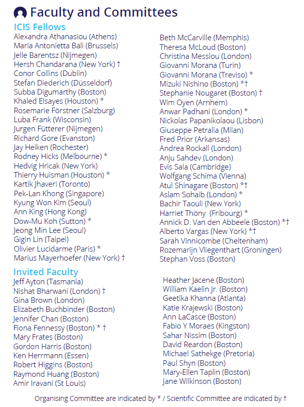
Meeting president: Prof. Annick D. Van den Abbeele
Relevant Documents
ICIS 2022 ProgrammeLearning objectives
To improve your knowledge of:
• The practice of Cancer Imaging on a Global Level
• Fostering Inclusion, Diversity and Equity on a Global Level
• The impact of COVID-19 on Cancer Imaging
• State-of-The-Art Practical Applications of Multimodal Cancer Imaging in most Major Cancers
• Multidisciplinary Teams’ Perspectives and Expectations in the Management of Patients with Cancer
• Molecular Cancer Imaging and Theranostics
• Recent Advances in Cancer Therapies including Immunotherapy and their impact on Cancer Imaging
• Use of Artificial Intelligence in Cancer Imaging
• Pediatric Cancer Imaging
• Cancer Genomics
• Image-guided Interventions
• Whole-Body MRI
• Tumor Imaging Metrics in Response Assessment
High quality educational lectures and hands-on workshops
• Refresh your knowledge of cancer imaging and learn about cutting edge developments
• Listen to state of the art multimodality cancer imaging (MRI, CT and PET)
• Improve your knowledge of the mechanism of different drugs in the treatment of cancer
• Bring your knowledge into practice in workstation based teaching in our hands-on workshops
• Experience the role of cancer imaging in multidisciplinary interaction: treatment selection, early response prediction and treatment adaptation
• Proffered papers (oral, poster and case presentations)

Cancer Imaging: An All-Inclusive Specialty
Venue: Hotel Commonwealth, 500 Commonwealth Avenue, Boston, 02215, MA. USA
Date: Monday 12th to Wednesday 14th September 2022
Keynote lectures
• A Keynote Lecture by Nobel Prize Laureate William G. Kaelin Jr., MD
• Fostering Diversity, Equity and Inclusion on a Global Level
• A Global View of The Lancet Oncology Commission on Medical Imaging and Nuclear Medicine by Prof. Hedvig Hricak
Main sessions
• Global Cancer Imaging: perspectives from each of the seven continents
• State-of-the-Art: Artificial Intelligence and Cancer Imaging
• Practical Cancer Imaging: Lung and Breast
• Brain and Head and Neck Cancer
• Liver Cancer
• Molecular Cancer Imaging and Theranostics
• State-of-the-Art Cancer Imaging: Pediatric Cancer Imaging, Genomics, Tumor markers
• State-of-the-Art: Immuno-oncology and Cancer Imaging
• Multidisciplinary Teams Perspectives and Expectations
• Practical Cancer Imaging: Pancreatic and Colorectal
Break-out sessions
• Cancer Imaging during the COVID-19 pandemic and Screening Incidental Chest Findings
• Practical Cancer Imaging: Kidney/Bladder/Prostate/Retroperitoneum and Gynecology Oncology
• Tumor Response Assessments
• Molecular Cancer Imaging and Theranostics
• Image-guided interventions
• State-of-the-Art Cancer Imaging: Pediatric Cancer Imaging and Genomics
Computer hands-on workshops Smaller classes, offered on a first come first served basis.
• Whole-body MRI
• Challenging prostate cancer cases and prostate MRI quality assessment
• Challenging renal and bladder oncology cases
• Challenging gynecology oncology cases
• Challenging brain and head/neck oncology cases
• Practice-oriented Cancer Imaging AI technology applications for clinical use
• Thyroid nodules: The role of TI-RADS
• LI-RADS made easy
• Challenging liver and pancreatic oncology cases
Scientific paper sessions and poster exhibition
Prizes for the best oral presentations and best posters will be awarded at the course party on Tuesday evening.
ICIS 2022 – Welcome Reception at the Hotel Commonwealth
A Welcome Reception will be held. Please join us for a drink and canapes.
A complimentary Welcome Reception with drinks and canapes hosted by ICIS President, Prof. Annick D. Van den Abbeele, will take place on Monday 12 September at the Hotel Commonwealth at the close of lectures. Please join us to enjoy an end of day drink with your fellow delegates, our sponsors and the ICIS 2022 Faculty.

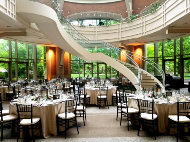
ICIS 2022 – Course Dinner
The Course Dinner will be held on Tuesday 13 September at Morton’s Boston Seaport (2 Seaport Lane, Boston, MA 02210).
Your ticket will include a courtesy bus to transport you from the conference venue to Morton’s, which is located in the very popular Seaport area of Boston, adjacent to the waterfront. You will have the opportunity to relax and network with faculty and other participants of ICIS 2022 as you enjoy a three-course meal with drinks in the attractive Atrium area of the restaurant. There will be a choice of entrees on the night; dietary requirements can be catered for.
Dinner tickets are available to purchase with registration at a cost of $95 per head; please be sure to indicate any dietary restrictions.
We hope you will join us at the dinner!
Entry requirements for USA
Closest Airport: Boston Logan
General Edward Lawrence Logan International Airport, also known as Logan International Airport is located in East Boston.
Onward travel from Logan
Water Shuttles: Connects with Downtown Boston and other interesting places. 66 route Shuttle bus for free between Boston Dock and all the terminals at the airport. See more information
MBTA: Public transportation offers different options to get from and to Logan Airport. See more information
Uber and Taxi: In all terminals, taxis wait outside the arrivals level. Fare to Downtown Boston fare is approximately $25-$45, depending on traffic / time of day. See more information
 Royal College of Radiologists – delegates will receive 18 credits in total for the three-day course. Monday 6.5 credits, Tuesday 6.5 credits, Wednesday 5 credits.
 The ICIS 2022: The 21st International Cancer Imaging Society Annual Teaching Course Cancer Imaging: An All-Inclusive Specialty is an Accredited Group Learning Activity (Section 1) as defined by the Maintenance of Certification (MOC) program of the Royal College of Physicians and Surgeons of Canada (Royal College) and has been approved by the Canadian Association of Radiologists (CAR) for a maximum of 18.0 credit hours.
Through an agreement between the Royal College and the American Medical Association, physicians may convert Royal College MOC credits to AMA PRA Category 1 Credits™. Information on the process to convert Royal College MOC credit to AMA credit can be found at www.ama-assn.org/go/internationalcme. Physicians should only claim credits commensurate with the extent of their participation in the activity.
Live educational activities recognized by the Royal College of Physicians and Surgeons of Canada as Accredited Group Learning Activities (Section 1) are deemed by the European Union of Medical Specialists (UEMS) eligible for ECMEC®
CPD certificates will be issued electronically, to the email address used to register for ICIS 2022. ICIS Members can download their CPD certificate in the Members' Area of the website, via their personal dashboard.
Submission deadline: Monday 1 June 2022
Guidelines
You may choose among four different abstract types:
> Oral Scientific Presentation
> Oral Case-Based Presentation
> Poster Scientific Presentation
> Poster Educational Presentation
Oral Scientific Presentation
The abstract should be separated into “Aim”, “Methods”, “Results” and “Conclusion”. The abstract limit is 250 words. Abstracts should not include promissory notes such as “We will provide additional data during our presentation.” Authors of accepted oral presentations will be invited for an online presentation within the Scientific Sessions. Presentation time will be 8 minutes with 2 minutes for Q&A (depending on the final program).
Oral Case-Based Presentation
The abstract should have an image and three teaching points. The abstract limit is 250 words. Abstracts should not include promissory notes such as “We will provide additional data during our presentation.” Authors of accepted oral case-based presentations will be invited for an online presentation within the Scientific Sessions. Presentation time will be 8 minutes with 2 minutes for Q&A (depending on the final program).
Poster Scientific Presentation
The abstract should be separated into “Aim”, “Methods”, “Results” and “Conclusion”. The abstract limit is 250 words.
Poster Educational Presentation
The abstract should be separated into “Learning Objectives”, “Content Organisation”, and “Conclusion”. The abstract limit is 250 words.
Abstracts that do not adhere to the following important points will be rejected:
> The title should not exceed 15 words. The title should be in bold, sentence case with no full stop at the end and no underlining.
> The abstract should not exceed 250 words.
> Please use authors’ initials and surnames only. No full stop at the end. Underline the name of the corresponding author. A comma should separate author names. Where authors are from a number of different institutions, the appropriate institution number from the affiliation list should be given as a superscript number immediately after each author's name, e.g.: John Smith1, Susan Jones2, Bill Fisher3. An asterisk * should be used to link the corresponding author with their email address
> Affiliations should include institute, town and country. Where there are multiple affiliations, each should be listed as a separate paragraph. Each institute should appear in the order used against the author names (see above paragraph) and show the appropriate superscript number, e.g.:
1 University, Town, State, USA
2 University, Town, UK
3 Company, Town, Country
> Qualifications should be omitted.
> Do not include references, tables or figures.
> Please do not use block capitals.
Main text
In structured abstracts, paragraph headings should be typed in bold with no colon at the end. Do not use the heading ‘Abstract’. Each heading should be in a separate paragraph.
Aim
Followed by regular text, on a new line and in the same format as shown above for main text.
Materials & Methods
Results
Conclusions
Consent to publish If the abstract contains details relating to individual participants (for example a case report), written informed consent for the publication of these details must be obtained from the participants and a statement to this effect should appear at the end of the abstract. Our guidelines for consent statements can be found here: http://www.biomedcentral.com/about/editorialpolicies#Ethics.
If the patient is deceased consent for publication should be obtained from the next of kin and if the patient is under 16 consent should be obtained from the parent or guardian.
Please find examples below
Proceedings of the 21st International Cancer Imaging Society Annual Teaching Course
Abstracts selected for presentation will be published in the Proceedings of the 21st International Cancer Imaging Society Annual Teaching Course, which will be made available online for all faculty and delegates. A downloadable version of your submission will be made available to our membership on the members only area of our website, and will be available on the Cancer Imaging open access website. Authors of selected abstracts will be notified by 1st July 2022.
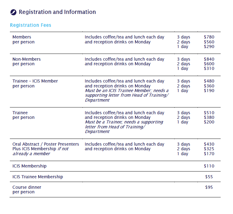
It will be obligatory for all scientific presenters to be members of ICIS at the time of online presentation. The annual membership fee of $110 will be added to the scientific presenters’ fee at registration if current membership is not in place. Please note that we have a discounted trainee membership at $55 for those who send a letter from their Head of Department, confirming their trainee status. Membership will run for one year from date of registration; all standard member benefits will apply.
Financial help will be available for junior proffered presenters. This will be decided on application and on a case-by-case basis.
Applicants for financial aid please email [email protected] for further information.
Queries may be addressed to the ICIS Secretariat by email to: [email protected]
EXAMPLE – ORAL CASE BASED PRESENTATION
Diagnosis: Advanced Cervix Cancer
Sala E.
Memorial Sloan Kettering Cancer Center, New York, USA
Images:
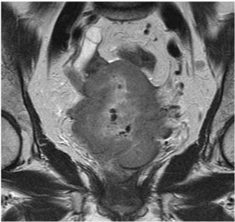
Teaching/Discussion points:
> Discuss the role of MRI in treatment slection and planning of patients with cervical cancer
> Describe MRI Features of parametrial and pelvic sidewall invasion
> Highlight potential pitfalls
EXAMPLE - POSTER EDUCATIONAL
Chemotherapy induced cardiomyopathy: an overview, imaging features, and future prospective
Firstname A Lastname1*, Firstname B Lastname2, Firstname C Lastname3
1University, Town, State, USA
2University, Town, UK
3Company, Town, Canada
*Email address of corresponding author if being included
Learning objectives:
To review the spectrum of imaging findings of chemotherapy- induced cardiomyopathy in correlation with most common cytotoxic drugs and regimens.
Content organisation:
Cardio toxic effect of chemotherapy is a well-recognized problem in cancer patients. Cardio toxicity depends on multiple predisposing factors, specific components of the chemotherapy regimen, length of treatment, and dosage.
We will present the spectrum of most common cardiotoxic chemotherapy agents and their combinations, specific effects on the myocardium, and imaging features of cardiomyopathies induced by chemotherapy.
We will review pathophysiology of chemotherapy induced cardiomyopathy including:
- Dose dependent cardiomyopathy
- Predisposing conditions –diabetes, presence of coronary artery disease, age.
- Potential reversibility
We will discuss imaging characteristics of chemotherapy induced cardiomyopathy
- Imaging modalities ( Echocardiography, Cardiac MR, and MUGA)
- Importance of monitoring cardiac function during and after treatment
- Distribution of late Gadolinium enhancement (LGE)
- Emerging technologies for early diagnosis of cardiomyopathy in cancer patients
Conclusions:
Chemotherapy induced cardiomyopathy is a common problem among cancer patients, increasing long term morbidity and mortality and often leading to disability. Patients receiving chemotherapy treatment, particularly cardio toxic agents, should be routinely assessed for cardiac function to diagnose cardiomyopathy during the early phase of treatment and to prevent development of irreversible heart failure.
EXAMPLE POSTER SCIENTIFIC
The value of 68Ga-PSMA enhanced MR-PET in patients with biochemical recurrent prostate cancer
Firstname A Lastname1*, Firstname B Lastname2, Firstname C Lastname3
1University, Town, State, USA
2University, Town, UK
3Company, Town, Canada
*Email address of corresponding author if being included
Aim: In patients with prostate Cancer increased levels of PSMA can be measured. Recently a new tracer, 68Ga-PSMA, was developed as a specific marker for hybrid imaging (PET/CT, MR-PET). In this study we evaluated the accuracy of 68Ga-PSMA in patients with rising PSA after radical prosatectomy, so called „biochemical recurrent prostate cancer“ (BRPC).
Materials and Methods: A total of 322 patients with BRPC underwent a MR-PET examination (Siemens Biograph mMR) after injection of about 150 mBq 68Ga-PSMA. Images were evaluated in cosensus by one experienced nuclear medicine physician and one radiologist. Pelvine lymphnode dissection was performed in most of the patients according to a predefined template with 8 fields. Lymphnode involvement was evaluated according to a 5 point scale with a patient- and a field-based analysis. These findings were startified according to PSA-values.
Results: Four patients were excluded from the study for different reasons. Sensitivity for detection of recurrence was 95.7 % for PSA-values ≥ 2ng/ml, 81.4 % for PSA-values of 1-2 ng/ml, 76% for PSA-values 0.5-1 ng/ml, and 51% for PSA values ≤ 0.5 ng/ml. In comparison to the MR-images alone MR-PET was of superior diagnostic value.
Conclusions: MR-PET using 68Ga-PSMA is a sensitive and highly accurate technique for the diagnosis of biochemical reccurence of prostate cancer after radical prostatectomy. It yields high diagnostic performance at relatively low PCA-values.
|
Don’t take our word for it. Read what our delegates said about our previous annual teaching course: • Very informative course for up to date knowledge on complex oncological imaging. |
Applications are invited for sponsorship to cover registration fees to participate in ICIS 2022, 12-14 September 2022, in Boston, MA. ICIS 2022 will be the 21st Annual Teaching Course of our Society and the first to be held in the USA. With the theme ‘Cancer Imaging: An All-Inclusive Specialty’, the three-day programme will include the perspectives of imaging from all seven continents of the world!
We would like to have a sponsored delegate representing as many of the continents as possible, to share this opportunity globally and therefore we welcome applications to secure sponsorship to join us for ICIS 2022!
Please email [email protected] with a description of how participating in ICIS 2022 will benefit your practice; entitle your email ‘ICIS 2022 sponsorship application’ and include a copy of your CV. Trainees should also submit a letter of support from their Head of Department.
The deadline for applications is Monday 4 July 2022.
You will be notified by 11 July 2022 whether or not your application has been successful, and whether support will cover virtual participation or attendance in-person at the three-day course.
|
Meeting Sponsors
Platinum sponsor: Bracco 
Gold sponsor: GE 
Silver sponsors: Guerbet, Lantheus, PETNET Solutions Inc., Siemens Healthineers Bronze sponsors: AI Metrics, Ashvattha Therapeutics, Centre for Probe Development & Commercialization, Hermes Medical Solutions, Hologic, IBA RadioPharma Solutions, ImaginAb, MIM Software Inc., Mint Medical, Visage Imaging. 


Supported by a grant from Pfizer Inc. |