Events / Education
22nd International Cancer Imaging Society Meeting and Annual Teaching Course
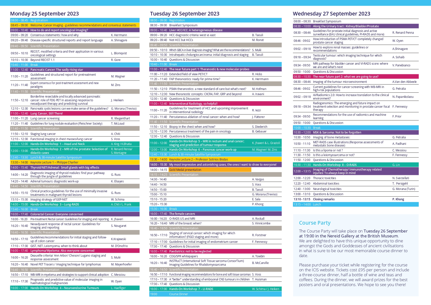
Mon 25 Sep 2023 Wed 27 Sep 2023 Society
IMPORTANT INFORMATION
Please complete all registration details. Registration will not be confirmed until full payment has been received.
Reservations are subject to availability and will be confirmed within one week from receipt of completed booking form and full payment. Please refer to our Booking Terms & Conditions.
No refunds will be made after 25 August 2023.
Please direct any questions to Louise Mustoe at [email protected]
Submission deadline: Monday 5 June 2023
Guidelines
You may choose among four different abstract types:
> Oral Scientific Presentation
> Oral Case-Based Presentation
> Poster Scientific Presentation
> Poster Educational Presentation
Oral Scientific Presentation
The abstract should be separated into “Aim”, “Methods”, “Results” and “Conclusion”. The abstract limit is 250 words. Abstracts should not include promissory notes such as “We will provide additional data during our presentation.” Authors of accepted oral presentations will be invited for an online presentation within the Scientific Sessions. Presentation time will be 8 minutes with 2 minutes for Q&A (depending on the final program).
Oral Case-Based Presentation
The abstract should have an image and three teaching points. The abstract limit is 250 words. Abstracts should not include promissory notes such as “We will provide additional data during our presentation.” Authors of accepted oral case-based presentations will be invited for an online presentation within the Scientific Sessions. Presentation time will be 8 minutes with 2 minutes for Q&A (depending on the final program).
Poster Scientific Presentation
The abstract should be separated into “Aim”, “Methods”, “Results” and “Conclusion”. The abstract limit is 250 words.
Poster Educational Presentation
The abstract should be separated into “Learning Objectives”, “Content Organisation”, and “Conclusion”. The abstract limit is 250 words.
Abstracts that do not adhere to the following important points will be rejected:
> The title should not exceed 15 words. The title should be in bold, sentence case with no full stop at the end and no underlining.
> The abstract should not exceed 250 words.
> Please use authors’ initials and surnames only. No full stop at the end. Underline the name of the corresponding author. A comma should separate author names. Where authors are from a number of different institutions, the appropriate institution number from the affiliation list should be given as a superscript number immediately after each author's name, e.g.: John Smith1, Susan Jones2, Bill Fisher3. An asterisk * should be used to link the corresponding author with their email address
> Affiliations should include institute, town and country. Where there are multiple affiliations, each should be listed as a separate paragraph. Each institute should appear in the order used against the author names (see above paragraph) and show the appropriate superscript number, e.g.:
1 University, Town, State, USA
2 University, Town, UK
3 Company, Town, Country
> Qualifications should be omitted.
> Do not include references, tables or figures.
> Please do not use block capitals.
Main text
In structured abstracts, paragraph headings should be typed in bold with no colon at the end. Do not use the heading ‘Abstract’. Each heading should be in a separate paragraph.
Aim
Followed by regular text, on a new line and in the same format as shown above for main text.
Materials & Methods
Results
Conclusions
Consent to publish If the abstract contains details relating to individual participants (for example a case report), written informed consent for the publication of these details must be obtained from the participants and a statement to this effect should appear at the end of the abstract. Our guidelines for consent statements can be found here: http://www.biomedcentral.com/about/editorialpolicies#Ethics.
If the patient is deceased consent for publication should be obtained from the next of kin and if the patient is under 16 consent should be obtained from the parent or guardian.
Please find examples below
Proceedings of the 21st International Cancer Imaging Society Annual Teaching Course
Abstracts selected for presentation will be published in the Proceedings of the 21st International Cancer Imaging Society Annual Teaching Course, which will be made available online for all faculty and delegates. A downloadable version of your submission will be made available to our membership on the members only area of our website, and will be available on the Cancer Imaging open access website. Authors of selected abstracts will be notified by 1st July 2022.
It will be obligatory for all scientific presenters to be members of ICIS at the time of online presentation. The annual membership fee of $110 will be added to the scientific presenters’ fee at registration if current membership is not in place. Please note that we have a discounted trainee membership at $55 for those who send a letter from their Head of Department, confirming their trainee status. Membership will run for one year from date of registration; all standard member benefits will apply.
Financial help will be available for junior proffered presenters. This will be decided on application and on a case-by-case basis.
Applicants for financial aid please email [email protected] for further information.
Queries may be addressed to the ICIS Secretariat by email to: [email protected]
EXAMPLE – ORAL CASE BASED PRESENTATION
Diagnosis: Advanced Cervix Cancer
Sala E.
Memorial Sloan Kettering Cancer Center, New York, USA
Images:
Teaching/Discussion points:
> Discuss the role of MRI in treatment slection and planning of patients with cervical cancer
> Describe MRI Features of parametrial and pelvic sidewall invasion
> Highlight potential pitfalls
EXAMPLE - POSTER EDUCATIONAL
Chemotherapy induced cardiomyopathy: an overview, imaging features, and future prospective
Firstname A Lastname1*, Firstname B Lastname2, Firstname C Lastname3
1University, Town, State, USA
2University, Town, UK
3Company, Town, Canada
*Email address of corresponding author if being included
Learning objectives:
To review the spectrum of imaging findings of chemotherapy- induced cardiomyopathy in correlation with most common cytotoxic drugs and regimens.
Content organisation:
Cardio toxic effect of chemotherapy is a well-recognized problem in cancer patients. Cardio toxicity depends on multiple predisposing factors, specific components of the chemotherapy regimen, length of treatment, and dosage.
We will present the spectrum of most common cardiotoxic chemotherapy agents and their combinations, specific effects on the myocardium, and imaging features of cardiomyopathies induced by chemotherapy.
We will review pathophysiology of chemotherapy induced cardiomyopathy including:
- Dose dependent cardiomyopathy
- Predisposing conditions –diabetes, presence of coronary artery disease, age.
- Potential reversibility
We will discuss imaging characteristics of chemotherapy induced cardiomyopathy
- Imaging modalities ( Echocardiography, Cardiac MR, and MUGA)
- Importance of monitoring cardiac function during and after treatment
- Distribution of late Gadolinium enhancement (LGE)
- Emerging technologies for early diagnosis of cardiomyopathy in cancer patients
Conclusions:
Chemotherapy induced cardiomyopathy is a common problem among cancer patients, increasing long term morbidity and mortality and often leading to disability. Patients receiving chemotherapy treatment, particularly cardio toxic agents, should be routinely assessed for cardiac function to diagnose cardiomyopathy during the early phase of treatment and to prevent development of irreversible heart failure.
EXAMPLE POSTER SCIENTIFIC
The value of 68Ga-PSMA enhanced MR-PET in patients with biochemical recurrent prostate cancer
Firstname A Lastname1*, Firstname B Lastname2, Firstname C Lastname3
1University, Town, State, USA
2University, Town, UK
3Company, Town, Canada
*Email address of corresponding author if being included
Aim: In patients with prostate Cancer increased levels of PSMA can be measured. Recently a new tracer, 68Ga-PSMA, was developed as a specific marker for hybrid imaging (PET/CT, MR-PET). In this study we evaluated the accuracy of 68Ga-PSMA in patients with rising PSA after radical prosatectomy, so called „biochemical recurrent prostate cancer“ (BRPC).
Materials and Methods: A total of 322 patients with BRPC underwent a MR-PET examination (Siemens Biograph mMR) after injection of about 150 mBq 68Ga-PSMA. Images were evaluated in cosensus by one experienced nuclear medicine physician and one radiologist. Pelvine lymphnode dissection was performed in most of the patients according to a predefined template with 8 fields. Lymphnode involvement was evaluated according to a 5 point scale with a patient- and a field-based analysis. These findings were startified according to PSA-values.
Results: Four patients were excluded from the study for different reasons. Sensitivity for detection of recurrence was 95.7 % for PSA-values ≥ 2ng/ml, 81.4 % for PSA-values of 1-2 ng/ml, 76% for PSA-values 0.5-1 ng/ml, and 51% for PSA values ≤ 0.5 ng/ml. In comparison to the MR-images alone MR-PET was of superior diagnostic value.
Conclusions: MR-PET using 68Ga-PSMA is a sensitive and highly accurate technique for the diagnosis of biochemical reccurence of prostate cancer after radical prostatectomy. It yields high diagnostic performance at relatively low PCA-values.
|
Don’t take our word for it; read what our delegates said about our previous annual teaching course:
• Very informative course for up to date knowledge on complex oncological imaging.
• Incredible conference with a really friendly atmosphere. The workshops give you a real insight into how to implement the techniques in practice.
• I strongly recommend this fantastic course.
• Excellent for learning, updating and inspiration to the challenges in your daily practice and also a very social meeting.
• The best Oncology Imaging meeting out there. This is a must-attend meeting for anyone interested in oncology related imaging.
• Excellent and productive course.
• Inspiring meeting.
• Great! Covers topics general radiologists need to know and do not easily find elsewhere.
• This was a highly informative conference in which it was easy to speak with experts in all fields informally at the breaks. I would highly
recommend it.
• Excellent. Strong case-based lectures with excellent literature references.
• Excellent speakers, great topics and great people, what more can you ask?
• Great conference with the latest cancer imaging information including criteria, protocol, modalities etc. Great information for the clinical trial in oncology using imaging. Amazing!
• ICIS annual meeting is the place where you can keep learning and create new projects in order to continue with your education in Oncological Imaging.
• A very well organised excellent course with superb speakers.
• Excellent cancer imaging forum that meets practice, academic and research needs. I have attended 14th, 15th, 16th and 18th Annual Teaching Courses. My knowledge has grown and I have shared this with colleagues from all over the world.
• Very good course. Highly recommended.
|
|
Meeting Sponsors
ICIS wish to thank the following companies who are supporting the 22nd Society Meeting and Annual Teaching Course: Gold Level: .png "GE Healthcare")

Bronze Level: 


|